About the shoulder muscles
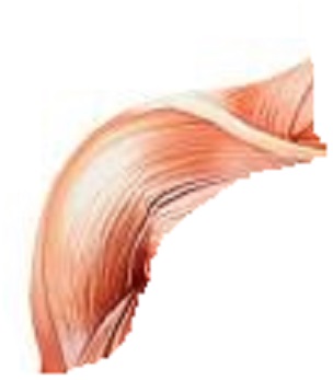
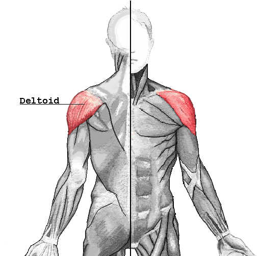
Deltoid
The deltoid muscle is the muscle forming the rounded contour of the human shoulder. It is also known as the 'common shoulder muscle', particularly in other animals such as the domestic cat. Anatomically, it appears to be made up of three distinct sets of fibers, namely 1. anterior or clavicular part (pars clavicularis) 2. posterior or scapular part (pars scapularis) 3. intermediate or acromial part (pars acromialis). However, electromyography suggests that it consists of at least seven groups that can be independently coordinated by the nervous system. It was previously called the deltoideus (plural deltoidei) and the name is still used by some anatomists. It is called so because it is in the shape of the Greek capital letter delta (Δ). Deltoid is also further shortened in slang as "delt".
Then all its fibers contract simultaneously, the deltoid is the prime mover of arm abduction along the frontal plane. The arm must be medially rotated for the deltoid to have maximum effect.[22] This makes the deltoid an antagonist muscle of the pectoralis major and latissimus dorsi during arm adduction. The anterior fibers assist the pectoralis major to flex the shoulder. The anterior deltoid also works in tandem with the subscapularis, pecs and lats to internally (medially) rotate the humerus.
The intermediate fibers perform basic shoulder abduction when the shoulder is internally rotated, and perform shoulder transverse abduction when the shoulder is externally rotated. They are not utilized significantly during strict transverse extension (shoulder internally rotated) such as in rowing movements, which use the posterior fibers.
The posterior fibers assist the latissimus dorsi to extend the shoulder. Other transverse extensors, the infraspinatus and teres minor, also work in tandem with the posterior deltoid as external (lateral) rotators, antagonists to strong internal rotators like the pecs and lats.
An important function of the deltoid in humans is preventing the dislocation of the humeral head when a person carries heavy loads. The function of abduction also means that it would help keep carried objects a safer distance away from the thighs to avoid hitting them, as during a farmer's walk. It also ensures a precise and rapid movement of the glenohumeral joint needed for hand and arm manipulation.[2] The intermediate fibers are in the most efficient position to perform this role, though like basic abduction movements (such as lateral raise) it is assisted by simultaneous co-contraction of anterior/posterior fibers.
The deltoid is responsible for elevating the arm in the scapular plane and its contraction in doing this also elevates the humeral head. To stop this compressing against the undersurface of the acromion the humeral head and injuring the supraspinatus tendon, there is a simultaneous contraction of some of the muscles of the rotator cuff: the infraspinatus and subscapularis primarily perform this role. In spite of this there may be still a 1–3 mm upward movement of the head of the humerus during the first 30° to 60° of arm elevation.
Source
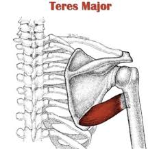
Teres Major
The teres major muscle is a muscle of the upper limb. It attaches to the scapula and the humerus and is one of the seven scapulohumeral muscles. It is a thick but somewhat flattened muscle. The teres major muscle (from Latin teres, meaning "rounded") is positioned above the latissimus dorsi muscle and assists in the extension and medial rotation of the humerus. This muscle is commonly confused as a rotator cuff muscle, but it is not because it does not attach to the capsule of the shoulder joint, unlike the teres minor muscle for example.
The teres major is a medial rotator and adductor of the humerus and assists the latissimus dorsi in drawing the previously raised humerus downwards and backwards (extension, but not hyperextension). It also helps stabilise the humeral head in the glenoid cavity.
Source
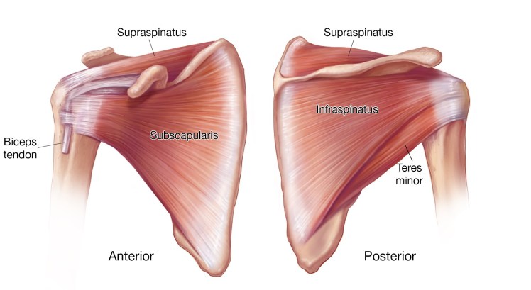
Supraspinatus
The supraspinatus (plural supraspinati) is a relatively small muscle of the upper back that runs from the supraspinous fossa superior portion of the scapula (shoulder blade) to the greater tubercle of the humerus. It is one of the four rotator cuff muscles and also abducts the arm at the shoulder. The spine of the scapula separates the supraspinatus muscle from the infraspinatus muscle, which originates below the spine.
The supraspinatus muscle performs abduction of the arm, and pulls the head of the humerus medially towards the glenoid cavity.[5] It independently prevents the head of the humerus to slip inferiorly.[5] The supraspinatus works in cooperation with the deltoid muscle to perform abduction, including when the arm is in adducted position.[5] Beyond 15 degrees the deltoid muscle becomes increasingly more effective at abducting the arm and becomes the main propagator of this action.
Source
Infraspinatus
In human anatomy, the infraspinatus muscle is a thick triangular muscle, which occupies the chief part of the infraspinatous fossa.[1] As one of the four muscles of the rotator cuff, the main function of the infraspinatus is to externally rotate the humerus and stabilize the shoulder joint.
The infraspinatus is the main external rotator of the shoulder. When the arm is fixed, it adducts the inferior angle of the scapula. Its synergists are teres minor and the deltoid. The infraspinatus and teres minor rotate the head of the humerus outward (external, or lateral, rotation); they also assist in carrying the arm backward. Additionally, the infraspinatus reinforces the capsule of the shoulder joint.
Source
Subscapularis
The subscapularis is a large triangular muscle which fills the subscapular fossa and inserts into the lesser tubercle of the humerus and the front of the capsule of the shoulder-joint. The subscapularis rotates the head of the humerus medially (internal rotation) and adducts it; when the arm is raised, it draws the humerus forward and downward. It is a powerful defense to the front of the shoulder-joint, preventing displacement of the head of the humerus.
Source
Teres Minor
The teres minor (Latin teres meaning 'rounded') is a narrow, elongated muscle of the rotator cuff. The muscle originates from the lateral border and adjacent posterior surface of the corresponding right or left scapula and inserts at both the greater tubercle of the humerus and the posterior surface of the joint capsule. The primary function of the teres minor is to modulate the action of the deltoid, preventing the humeral head from sliding upward as the arm is abducted. It also functions to rotate the humerus laterally. The teres minor is innervated by the axillary nerve.
The infraspinatus and teres minor attach to head of the humerus; as part of the rotator cuff they help hold the humeral head in the glenoid cavity of the scapula. They work in tandem with the posterior deltoid to externally (laterally) rotate the humerus, as well as adduction. Teres Minor can produce only very small scapular plane adduction during maximal contraction (Hughes RE, An KN 1996) with adductor moment arm of approximately 0.2 cm at 45° of shoulder internal rotation and approximately 0.1 cm at 45° of shoulder external rotation.
Source
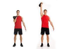
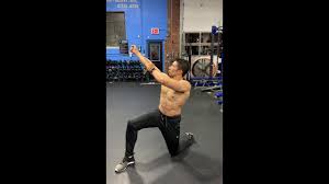
Exercises
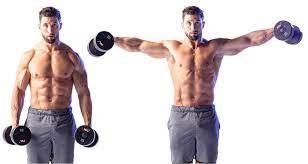
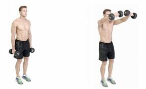
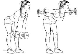
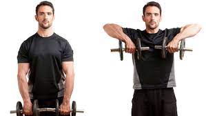
Stretches
- Across-the-chest stretch
- Bring your right arm across your chest.
- Place it in the crease of your left elbow or use your left hand to support your arm.
- Hold this position for up to 1 minute.
- Repeat on the opposite side.
- Neck Release
- Lower your chin toward your chest. You’ll feel a stretch along the back of your neck.
- Gently tilt your head to the left to stretch your right shoulder.
- Hold this position for up to 1 minute.
- Repeat on the opposite side.
- Chest Expansion
- While standing, hold an exercise band, strap, or towel behind your back with both hands.
- Broaden across your chest as your move your shoulder blades toward each other.
- Lift your chin and look up toward the ceiling.
- Hold for up to 30 seconds.
- Seated Twist
- Sit in a chair with your ankles directly under your knees.
- Twist your upper body to the right, bringing the back of your left hand to your thigh.
- Place your right hand down wherever it’s comfortable.
- Hold this position for up to 30 seconds.
- Repeat on the left side.
- Shoulder Circles
- Stand with your left hand on the back of a chair.
- Allow your right hand to hang down.
- Circle your right hand 5 times in each direction.
- Repeat on the opposite side.
- Thread the Needle
- Start on your hands and knees. Lift your right hand up toward the ceiling with your palm facing away from your body.
- Lower your arm to bring it under your chest and over to the left side of your body with your palm facing up.
- Activate your right shoulder and arm to avoid collapsing into this area.
- Keep your left hand on the floor for support, lift it toward the ceiling, or bring it around to the inside of your right thigh.
- Hold this position for up to 30 seconds.
Injuries
Most problems in the shoulder involve the muscles, ligaments, and tendons, rather than the bones. Athletes are especially susceptible to shoulder problems. In athletes, shoulder problems can develop slowly through repetitive, intensive training routines.Some people will have a tendency to ignore the pain and "play through" a shoulder injury, which only aggravates the condition, and may possibly cause more problems. People also may underestimate the extent of their injury because steady pain, weakness in the arm, or limitation of joint motion will become almost second nature to them.
Sometimes, one of the shoulder joints moves or is forced out of its normal position. This condition is called instability, and can result in a dislocation of one of the joints in the shoulder. Individuals suffering from an instability problem will experience pain when raising their arm. They also may feel as if their shoulder is slipping out of place.Impingement is caused by excessive rubbing of the shoulder muscles against the top part of the shoulder blade, called the acromion. Impingement problems can occur during activities that require excessive overhead arm motion. Medical care should be sought immediately for inflammation in the shoulder because it could eventually lead to a more serious injury.
The rotator cuff is one of the most important components of the shoulder. It is comprised of a group of muscles and tendons that hold the bones of the shoulder joint together. The rotator cuff muscles provide individuals with the ability to lift their arm and reach overhead. When the rotator cuff is injured, people sometimes do not recover the full shoulder function needed to properly participate in an athletic activity.
Source